提醒成功


Mouse Monoclonal Antibody to TDP43
| Description |
|---|
HIV-1, the causative agent of acquired immunodeficiency syndrome (AIDS), contains an RNA genome that produces a chromosomally integrated DNA during the replicative cycle. Activation of HIV-1 gene expression by the transactivator Tat is dependent on an RNA regulatory element (TAR) located downstream of the transcription initiation site. The protein encoded by this gene is a transcriptional repressor that binds to chromosomally integrated TAR DNA and represses HIV-1 transcription. In addition, this protein regulates alternate splicing of the CFTR gene. A similar pseudogene is present on chromosome 20. [provided by RefSeq, Jul 2008] |
| References |
|---|
| 1,Adv Exp Med Biol. 2021;1281:201-217. 2,Science. 2021 Feb 5;371(6529):eabb4309. |
| Specification | |
|---|---|
| Aliases | ALS10; TDP-43 |
| Entrez GeneID | 23435 |
| clone | 6G2D8 |
| WB Predicted band size | 44.7kDa |
| Host/Isotype | Mouse IgG1 |
| Antibody Type | Primary antibody |
| Storage | Store at 4°C short term. Aliquot and store at -20°C long term. Avoid freeze/thaw cycles. |
| Species Reactivity | Human |
| Immunogen | Purified recombinant fragment of human TDP43 (AA: free peptide) expressed in E. Coli. |
| Formulation | Purified antibody in PBS with 0.05% sodium azide |
| Application | |
|---|---|
| WB | 1/500 - 1/2000 |
| IHC | 1/200 - 1/1000 |
| ICC | 1/200 - 1/1000 |
| FCM | 1/200 - 1/400 |
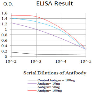
| ELISA | 1/10000 |
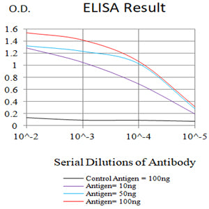
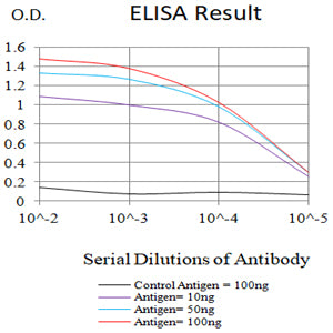
- Black line: Control Antigen (100 ng);Purple line: Antigen (10ng); Blue line: Antigen (50 ng); Red line:Antigen (100 ng)
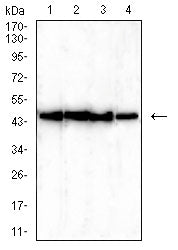
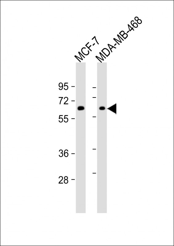
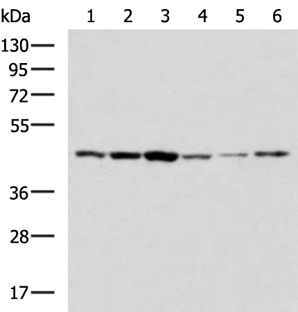
- Western blot analysis using TDP43 mouse mAb against Hela (1), HEK293 (2), MCF-7 (3), and A549 (4) cell lysate.
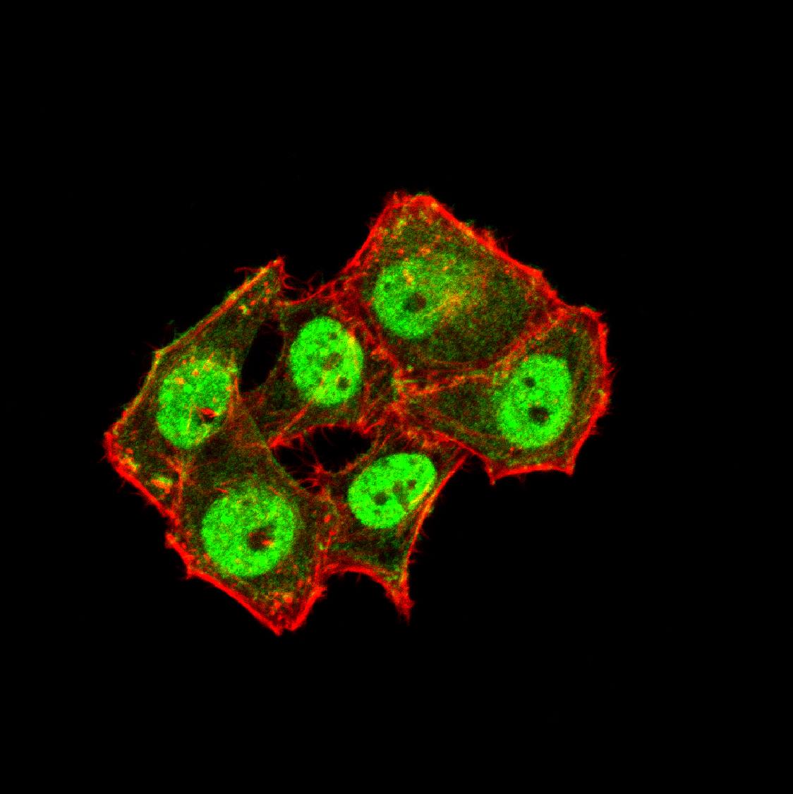
- Immunofluorescence analysis of Hela cells using TDP43 mouse mAb (green). Blue: DRAQ5 fluorescent DNA dye. Red: Actin filaments have been labeled with Alexa Fluor- 555 phalloidin. Secondary antibody from Fisher (Cat#: 35503)
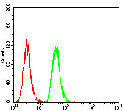
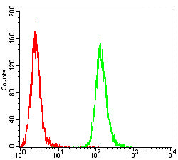
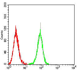
- Flow cytometric analysis of A431 cells using TDP43 mouse mAb (green) and negative control (red).
- Flow cytometric analysis of Hela cells using TDP43 mouse mAb (green) and negative control (red).
- Flow cytometric analysis of HepG2 cells using TDP43 mouse mAb (green) and negative control (red).
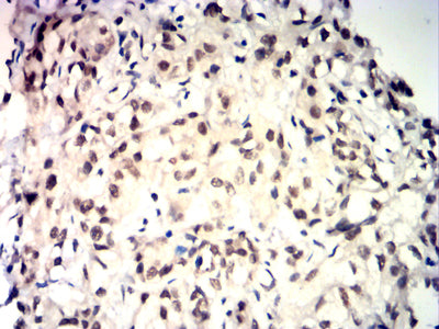
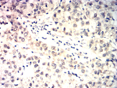
- Immunohistochemical analysis of paraffin-embedded human baldder cancer tissues using TDP43 mouse mAb with DAB staining.
- Immunohistochemical analysis of paraffin-embedded human liver cancer tissues using TDP43 mouse mAb with DAB staining.
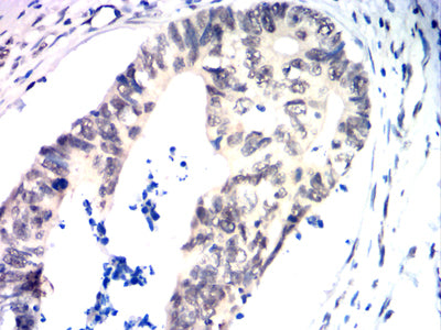
- Immunohistochemical analysis of paraffin-embedded human rectal cancer tissues using TDP43 mouse mAb with DAB staining.
For Reseach Only
Application Key:WB - Western Blot | IHC - Immunohistochemistry | ICC - Immunocytochemistry | FCM - Flow Cytometry | ELISA - Enzyme-linked Immunosorbent Assay | IP - Immunoprecipitation
#90009
相关产品


 微信/QQ登录


 首页
首页
